DISORDERS OF THE EAR Pt. 2
EDUCATION
Continuing our video-otoscope guide to the characteristics of disorders of the ear that audiologists should spot and provide referrals on. Part two of this review looks at visible signs of some serious conditions, introduced by expert Dr. Rakesh Chopra.

Professionals should, for example, be able to spot the lurking cholesteatoma or a malignant process behind presentation of hearing loss.
If you missed Part One of this two-part outline, catch it here.
About Dr. Chopra
Dr. Rakesh Chopra is a gifted lecturer, whose St. Helens-based ENT GPwER service is now in its 18th year. Prior to that, he had 14 years in hospital-based ENT. He has authored multiple international publications in the field, and does ENT triage for St. Helens, Warrington, and Knowsley. Outside of medicine, he has published three novels and is a musician.
“Sound is an amazing thing; we take it for granted,” Dr. Chopra told audiologists at the start of his October presentation to delegates at the annual conference of the British Society of Hearing Aid Audiologists (BSHAA). “As time is passing, we are knowing more and more about how important sound is for the human brain, much more than we thought” he continued, adding: “Music is called the physician of the future.”
Read on and take note of the video-otoscopic images.
1. Chronic suppurative otitis media
Tympanic Membrane perforations. It is important to ascertain where the hole in the eardrum is located. If it is in the bulk of the tympanic membrane – called the pars tensa, these are called central perforations and considered safe because they are not usually associated with serious complications. The problems are recurrent infections, ear discharge, and hearing loss.
Other perforations or retraction pockets occur in the area of the eardrum called the attic. These need to be managed differently as there is a risk of cholesteatoma formation, as well as potential for complications such as facial palsy or intracranial infections. These are considered unsafe. Marginal and attic perforations – as below examples of postero-superior pathology and attic disease – are unsafe.


2. Malignancy in the ear
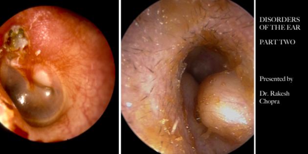
The picture shows (left side) a squamous cell carcinoma of the ear canal, compared with just dry skin (right side)!
3. Exostosis
Benign bony outgrowths in the proximal ear canal may be seen in people who do a lot of swimming. No treatment is required unless symptomatic.
4. Glomus Tumours
These are a rare benign vascular neoplasm called a glomus body, which arises from the neuro-arterial structure . They are paragangliomas, and may present with a rising sun appearance behind the eardrum. Pulsatile tinnitus and hearing loss are the common presentations.
5. A High riding Jugular Bulb
The jugular bulb sits in close proximity to the floor of the middle ear, and in some cases it can be invested higher. Its bony covering may be absent (4%) and can be seen behind the tympanic membrane. It can present with pulsatile tinnitus and an abnormal-looking eardrum.
Source: Audio Infos issue 152 January-February 2023


